Blue nevus
| Blue nevus | |
|---|---|
| Other names | Blue neuronevus, dermal melanocytoma, nevus coeruleus, nevus bleu[1] |
 | |
| Blue nevus | |
| Specialty | Dermatology |
| Symptoms | Single well-defined blue-black bump[2] |
| Complications | Rarely malignant transformation[3] |
| Types | Dendritic, cellular[2] |
| Causes | Unclear[3] |
| Diagnostic method | Visualisation, dermoscopy[4] |
| Differential diagnosis | Dermatofibroma, melanoma[3][5] |
| Treatment | Monitoring, excision[3] |
| Prognosis | Good[3] |
| Frequency | Female>male[2] |
A blue nevus is a type of coloured mole, typically a single well-defined blue-black bump.[1][2]
The blue colour is caused by the pigment being deep in the skin.[4]
Diagnosis is by visualisation and dermoscopy.[4] A biopsy is sometimes performed, or the whole lesion surgically removed.[3] The outcome is generally good but there is a small chance of cancerous transformation.[3] Differential diagnosis includes dermatofibroma and melanoma.[3]
Blue nevi are more common in females than males.[2] It was first studied in 1906 by Tièche, a student of Josef Jadassohn.[6]
Classification
Blue nevi may be divided into the following types:[7]: 701
- A patch blue nevus (also known as an "acquired dermal melanocytosis", and "dermal melanocyte hamartoma") is a cutaneous condition characterized by a diffusely gray-blue area that may have superimposed darker macules.[1]
- A blue nevus of Jadassohn–Tièche (also known as a "common blue nevus", and "nevus ceruleus") is a cutaneous condition characterized by a steel-blue papule or nodule.[7]: 701
- A cellular blue nevus is a cutaneous condition characterized by large, firm, blue or blue-black nodules.[7]: 701
- An epithelioid blue nevus is a cutaneous condition most commonly seen in patients with the Carney complex.[7]: 701
- A deep penetrating nevus is a type of benign melanocytic skin tumor characterized, as its name suggests, by penetration into the deep dermis and/or subcutis. Smudged chromatic is a typical finding. In some cases mitotic figures or atypical melanocytic cytology are seen, potentially mimicking a melanoma. Evaluation by an expert skin pathologist is advisable in some cases to help differentiate from invasive melanoma.[7]: 701
- An amelanotic blue nevus (also known as a "hypomelanotic blue nevus") is a cutaneous condition characterized by mild atypia and pleomorphism.[7]: 701
- A malignant blue nevus is a cutaneous condition characterized by a sheet-like growth pattern, mitoses, necrosis, and cellular atypia.[1][7]: 701
-
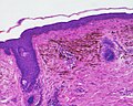
 Micrograph of a blue nevus showing the characteristic pigmented melanocytes between bundles of collagen. H&E stain
 -
 Blue nevus
 -
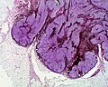 Cellular blue nevus
 -
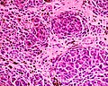 Epithelioid blue nevus
 -
 Malignant blue nevus


See also
References
- ^ a b c d Rapini, Ronald P.; Bolognia, Jean L.; Jorizzo, Joseph L. (2007). Dermatology: 2-Volume Set. St. Louis: Mosby. p. 1722. ISBN 978-1-4160-2999-1.
- ^ a b c d e Johnstone, Ronald B. (2017). "32. Lentigines and melanomas". Weedon's Skin Pathology Essentials (2nd ed.). Elsevier. p. 545. ISBN 978-0-7020-6830-0.
- ^ a b c d e f g h Austad, Steve S.; Athalye, Leela (2021). "Blue Nevus". StatPearls. StatPearls Publishing. PMID 31747181.
- ^ a b c "Blue naevus". dermnetnz.org. Retrieved 21 October 2021.
- ^ Blue+Nevi at the U.S. National Library of Medicine Medical Subject Headings (MeSH)
- ^ Sreeremya, S. (17 April 2018). "Blue Nevus". International Journal of Molecular Biotechnology. 4 (1): 1–4. doi:10.37628/ijmb.v4i1.255 (inactive 11 July 2025).
{{cite journal}}: CS1 maint: DOI inactive as of July 2025 (link) - ^ a b c d e f g James, William D.; Berger, Timothy G.; et al. (2006). Andrews' Diseases of the Skin: clinical Dermatology. Saunders Elsevier. ISBN 0-7216-2921-0.
External links
Content Disclaimer
Informasi ini disarikan dari Wikipedia dan disajikan kembali untuk tujuan edukasi. Konten tersedia di bawah lisensi CC BY-SA 3.0. Kami tidak bertanggung jawab atas ketidakakuratan data yang bersumber dari kontribusi publik tersebut.
- The information displayed on this website is sourced in part or in whole from Wikipedia and has been adapted for the purpose of restating it. We strive to provide accurate and relevant information, however:
- There is no guarantee of absolute accuracy. Wikipedia is an open, collaborative project that can be edited by anyone, so information is subject to change.
- It is not intended to constitute professional advice. The content displayed is for informational and educational purposes only. For important decisions (e.g., medical, legal, or financial), please consult a professional.
- Content copyright. Wikipedia is licensed under the Creative Commons Attribution-ShareAlike License (CC BY-SA). This means that content may be reused with appropriate attribution and shared under a similar license.
- Responsible use. Any risk arising from the use of information from this website is entirely the responsibility of the user.